sub-imageதேர்தல் களம்
sub-imageதினமலர் டிவி
sub-imagePodcast
sub-imageiPaper
sub-imageசினிமா
sub-imageகோயில்கள்
sub-imageபுத்தகங்கள்
sub-imageSubscription
sub-imageதிருக்குறள்
sub-imageகடல் தாமரை
Dinamalar Logo

வியாழன், மே 14, 2026 ,சித்திரை 31, பராபவ வருடம்

டைம்லைன்


தற்போதைய செய்தி


தினமலர் டிவி


ப்ரீமியம்


தமிழகம்


இந்தியா


உலகம்


வர்த்தகம்


விளையாட்டு


கல்விமலர்


டீ கடை பெஞ்ச்

தினம் தினம்


ஜோசியம்


காலண்டர்


ஆன்மிகம்


வாராவாரம்


இணைப்பு மலர்


போட்டோ


உலக தமிழர்


ஸ்பெஷல்


/வாராவாரம்/பட்டம்/First Ever Nanoscale Image of a Living Cell Membrane

First Ever Nanoscale Image of a Living Cell Membrane

First Ever Nanoscale Image of a Living Cell Membrane


PUBLISHED ON : ஜூன் 05, 2017

Follow on GoogleFavourite on Google

PUBLISHED ON : ஜூன் 05, 2017


Follow on GoogleFavourite on Google
Latest Tamil News
அ நிறம் | அளவு

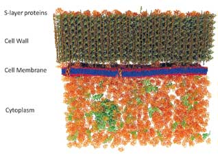
Researchers have completed the world's first scan of a living cell membrane down to a nanoscale level. Its details could finally resolve a longstanding debate on how they function.

The research was carried out by a team of scientists from Oak Ridge National Laboratory in Tennessee, who used a mix of genetic and chemical labelling techniques to add an isotope of hydrogen to the membranes of living Bacillus subtilis cells.

The technique used to create this incredible image could fundamentally change how nanoscale structures are studied in living things.

தினமலர் WhatsApp Channel-ல் சேருங்கள்
Latest Tamil news, instantly on WhatsApp
Follow


      Dinamalar
      Follow us